
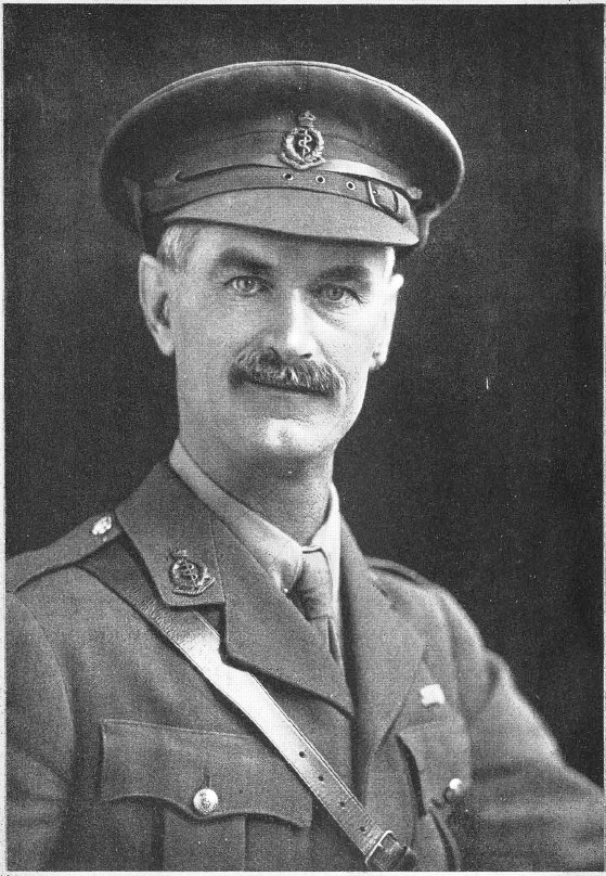
Not in so distant past, most of Kashmir lacked resources to even have adequate garments in winters. Kangri, the fire pot was the only warm tool but its overuse was triggering peculiar cancer endemic to Kashmir. In this piece, one of the two Neve brothers who dedicated their life to Kashmir, Arthur Neve details the causes, symptoms, prevalence, and treatment of this cancer in 1900. Though techniques in treating the disease and terminology used to describe the crisis have changed in the last 120 years the write-up is vital in understanding Kashmir’s life and times early last century
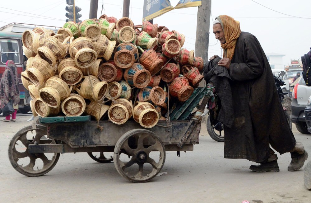
PHOTO BY BILAL BAHADUR
Epithelioma, arising on skin surfaces, other than the neighbourhood of mucous membrane, is of rather rare occurrence in England. But in Kashmir, the commonest seat of epitheliomata is the skin of the thigh and the abdomen.
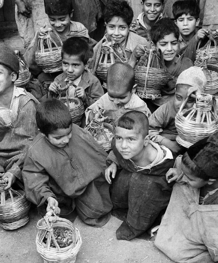
The cause of this is not far to seek. During the severe winters and indeed for a great part of the year, every Kashmiri man, woman or child carries a portable charcoal brazier under the loose gown which constitutes his or her only garment. When walking, this hangs against the abdomen, when seated it is placed on the ground between the thighs. Slight burns frequently occur, and in very many people there is a mottling of the skin in the exposed parts.
The frequency of epithelioma may be judged by the fact that twenty cases of this kind were operated upon during June and July only. Probably two or three inoperable cases were seen in the same period.
Age: It is difficult to ascertain the ages of our native patients, but it may be approximately stated that of the above, one was under forty, two under fifty, six under sixty, nine under seventy, two under eighty, and one above ninety.
Sex: Most of the cases are males, fifteen out of twenty.
Region: Nine occurred in the thigh, four on the abdomen, three on the lower part of the chest, two on the leg, one each on the back and hand.
The glands of the groin and axilla are often affected, and these constitute by far the gravest aspect of the disease.
Clinical appearances: There may have been an ulcer or warty growth preceding the epithelioma, and such are often seen side by side with it. In either case, the transformation to malignancy is indicated by a central ulceration, with foul discharge, an eversion and thickening of the edge.
In my last case, the ulcer was only half an inch in diameter.
Another recent case had a horny tumour an inch in diameter, conical in shape, one edge of which was undermined, and examination by Stiles’ method showed that the basement membrane was no longer intact, but the epithelial tissue had begun to invade the adipose tissue beneath. A special interest attaches to these marginal cases, which are not common, so it is curious that only a few days later another horny tumour was seen in which the horny tissue was lifted and partly broken up by a mass of epithelial tissue, which also infiltrated the deeper structures.
But the usual appearance is that of a foul, irregular, and somewhat raised ulcer, partly covered by sloughy detritus and crusts. It may appear as a cauliflower excrescence of dusky granulations or may be crater shaped.
When in the abdomen the excavation is likely to be deeper than when on the thigh, and when deep the glands are almost certain to be affected.
The disease is often central, in which case both groins are apt to be infected, but when the epithelioma is situated above the umbilicus it is the glands of the axilla not of the groin which will be the site of secondary growths. These gland tumours are usually brawny at quite an early period and tend soon to break down and infiltrate surrounding structures.
 Structure: This is identical with that of epithelioma occurring in other situations such as the lip or scrotum. The epithelium of the adjoining skin dips down, proliferates, forms the familiar cell-nests, is itself invaded by connective tissue cells, which may form granulations on the surface. Even when fungating there is little tendency to bleeding, as the vessels are not numerous, on the contrary, the smaller vessels are often occluded, and hence there is gangrene of superficial portions of the tumour. As the finger-like processes invade the deeper tissues they are surrounded and to some extent marked off from the healthy structures by connective tissue formation. It is comparatively seldom that the muscles of the thigh are involved, but in the abdomen, the tendinous and muscular tissues soon blend with the epitheliomatous, and the parietal peritoneum is sooner or later infiltrated.
In the chest too the early tendency to infiltrate is marked, and the periosteum of the ribs becomes involved, as in one of the cases under review.
As would be expected the secondary gland tumours occur most frequently in connection with epitheliomas of the ulcerative and infiltrating type, but the most harmless-looking warty epithelioma may, as in a recent case, be the starting point of an inoperable glandular tumour.
The infection is of course conveyed along the lymphatics, but it is seldom that the route can be traced by any line of shot-like deposits. And the rapid brawny infiltration sometimes met with in cases where the glandular growth is not broken down or very extensive is difficult to explain.
Secondary deposits are seldom confined to one or two glands. The groin glands are affected in connection with either thigh or abdominal tumours, but clinically. I have observed that the deeper glands within the abdominal cavity are more often enlarged in relation to thigh disease.
Treatment: This may be summed up by the word operation. Some are inoperable, and one occasionally employs caustics in such cases. And of all the caustics I have used I regard formalin as the most powerful, but in the cases now under review, the knife has been employed.
It is usually a comparatively simple thing to remove the original tumour. The edge is sharply defined by the ulceration, and when examined, whether by Stiles’ nitric acid method or by the microscope, it will be found that the epitheliomatous structure does not extend above a quarter of an inch beyond the apparent margin.
Care has to be taken to cut wide of the roots in fascia or muscles, but these are usually well defined. When situated on the thigh unless the diameter exceeds three inches it will be found possible by tension to unite the skin, but the stitches will often cut out. This is the more often because there is great septicity to start with.
Burning Cancers With Iron
I have endeavoured in some recent cases to get a cleaner field for operation by thoroughly cauterising the ulcerating mass under chloroform with a red-hot iron, and then scrubbing the surrounding parts with antiseptics. This is a practical point of great value in other than epithelioma cases. It may not be possible to make the skin sterile in a few minutes, but much may be done to reduce its septicity, and by care, the deeper portions of the wound may escape infection, in which case early union may save the subsequent breaking down of the skin incision.
Button sutures are useful also. In the abdominal wall, the constant respiratory movements are a further hindrance to immediate healing. It is usually easiest to unite the wound across the axis of the body, and a short splint or firm pad helps to restrain movement. A few vessels may require ligature, but when these are superficial, I prefer to put on forcipressure for a few seconds and then include the vessels in the skin sutures.
Portions of the gracilis or adductor muscles have occasionally to be removed. In the abdomen, the muscles tend to ulcerate away, and the peritoneum may be perilously near the surface and be wounded. This happened in one of my cases some years ago, and the tension on the sutures was such that firm union could not be obtained. It ended fatally by slow septic absorption from the peritoneal cavity. This has been the only fatal case on over three hundred operations for epithelioma performed by Dr E L Neve or myself since 1890.
No Complete Cure
But, as in other departments of surgery, the successful operation does not ensure a complete cure of disease. The great problem is not the removal of the original tumour but of the secondary glandular ones. Much has been written about the complete operation for scirrhus of the breast; Stiles and Watson Cheyne have done much to impress the immense importance of complete eradication of the outlying glandular tissue, and also of all the lymph glands of the axilla.
Here too we are faced by the same problem, but the difficulties are even greater. The superficial glands of the groin may indeed be as easy of excision as axillary glands – not so the deeper glands surrounding the femoral sheath. The common femoral itself is not infrequently involved, and there are iliac glands, which become infiltrated. The question is as to the legitimate boundaries of surgery.
Butlin maintains that the operative surgery of malignant disease has, of late years, been pushed beyond reasonable limits. One thing is clear, namely, that when the groin glands are already visibly and extensively implicated, and especially when there is any brawny infiltration of the skin, it is too late to achieve a radical cure, and any severe attempts in that direction are misplaced.
Recent Cases
Speaking from recent experience in two of the cases now under analysis, a sweeping operation has failed to give any relief but has appeared only to precipitate general dissemination of disease in surrounding tissues. One such is still in the wards, in whom I attempted to extirpate the groin disease, removing the skin freely, and dissecting out the whole of the subcutaneous fatty and glandular tissue en bloc over an area eight inches long and five inches wide.
I cleaned the sheath of the femoral vessels and did not suspect any source of direct epitheliomatous infection of the wound. Yet already the most widespread ulcerative disease is visible above and to the inside of the wound, which is nearly closed.
In another recent case, I removed all enlarged glands in both groins, secondary to an abdominal tumour, but within a few weeks, other large glands with diffuse hardness made their appearance. Cure can only be expected where the disease is not already generalised in the lymphatics of the region.
In some of the slow-growing papillomatous type, many months or a year may elapse before the glands are effected, and if early removed the groin may be left untouched. Fortunately, the least enlargement of the groin glands can be detected by careful examination.
In the case of the thigh, it is well to remove at least the superficial line of glands accompanying the saphenous vein, which will often be found thrombosed. And if one of these be found to react to the nitric acid method, it would then be well to proceed to clear the glands along the femoral sheath and those parallel to Poupart’s ligament. Great care has to be exercised not to diffuse epitheliomatous cells by rough handling of the glands, or with the knife smeared by the cancerous juice.
Of the 20 cases, there were glandular enlargements in six. It is doubtful whether any of these can be regarded as cured. There has certainly been a recurrence in four already. This is a considerably higher rate of recurrence than I had observed when writing on the subject ten years ago, perhaps because patients now return more readily for further operative treatment.
Numbers
The statistics for a considerable period are as follows: From 1881 to 1889 there were 169 cases, of these two died, seven were partial operations, with but temporary improvement; 160 were entered as cured.
From 1890 to 1898 inclusive, there have been 316 cases, of which one died, seven were but temporarily improved, and 310 have been entered as cured.
These figures do not give a true idea of the real mortality of the disease.
There are the other cases regarded when first seen as inoperable, and treated by palliatives or caustics. Then the recurrences not seen by us or not registered must be numerous. This may be fudged by the comparatively large number of gland operations during recent years.
Thus, in the last two years, out of 70 operations, there have been also 35 operations on the glands of the axilla or groin. Perhaps, in earlier years, many of these glands would have been left untouched. There is no reason to suppose that the type of disease is becoming more malignant.
To summarise our experience: Kangri-burn cancer is a squamous-celled epithelioma of the skin. It occurs only on surfaces constantly exposed to great heat and to occasional burns. Secondary gland infection is frequent, and is as malignant as other glandular epithelioma. In the early stage, the disease is localised, and a radical cure can be effected by free excision. The permanent cures may be as high as 50 per cent. When the glands of the groin are infected, complete removal becomes exceedingly difficult, and recurrences are frequent. It is inadvisable to do very sweeping operations where the glands are extensively involved or the skin over them brawny. The direct operation mortality on 485 cases has been only 6 per cent.
(This write-up appeared in The Indian Medical Gazette in March 1900 under the title Kangri-Burn Epithelioma In Kashmir)


